
Can We Help You?
x
In this issue:

How Laser Spectroscopy Advances Biotech
Spectroscopy may be broadly defined as the study of the interaction between matter and radiation. What makes spectroscopy truly interesting is that it�s arguably the most widely applied study method in modern science. Experimental spectroscopic studies have been central to the development of quantum mechanics, and they included the explanation of Blackbody Radiation, Photoelectric Effect and Niels Bohr's explanation of atomic structure and spectra. Since atoms and molecules have their own unique spectra response, spectroscopy can be used in Applied Chemistry to detect, identify and quantify information about particles and hence distinguish one type of matter from another. This fundamental benefit of spectroscopy has made it invaluable for Biotechnology and Life Sciences.
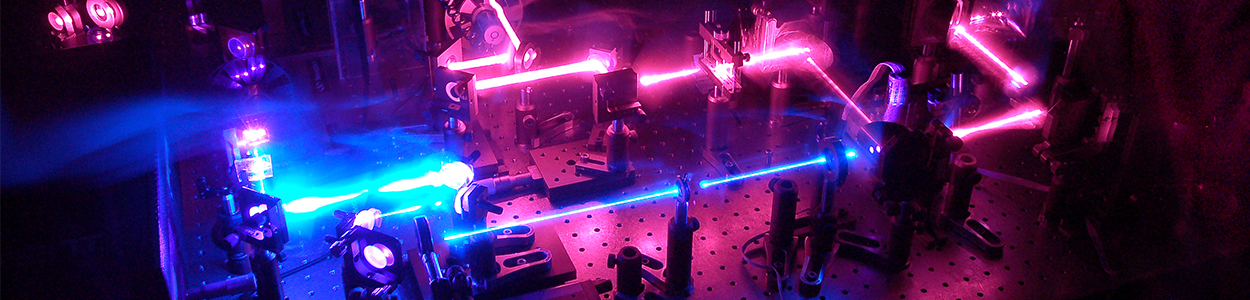
Expressed mathematically, spectroscopy refers to the measurement of radiation intensity as a function of wavelength. However, it can be divided into multiple types based on the type of radiative energy, and the nature of interaction between the matter and energy. In Laser Spectroscopy, researchers train a laser beam on a sample that yields a characteristic light which can be analyzed by a spectrometer. Laser Spectroscopy can in turn be divided into different types based on the type of laser used, and the atom's response that is studied.
In Laser-Induced-Breakdown Spectroscopy (LIB-S), a sample is excited with intense laser pulses and the emitted light is analyzed, which generally falls in the visible or infrared spectral region, i.e. having wavelengths longer than that of the pump source. LIBS is ideal for sample analysis as it doesn't require sample preparation, and generally removes less than 1 gram of material during laser ablation. In Raman Spectroscopy, a substance is irradiated with a narrow-band light at a high optical power to detect the weak emission lines which arise from spontaneous or stimulated Raman Scattering1. The spectrum of Raman Scattered light contains information about molecular vibrations, and helps identify key properties of the material - such as wavenumber scale for example, which is essential in pharma-applications.
The past few years have seen a surge in Biotech applications that use laser spectroscopy. Raman Spectroscopy in conjunction with dynamic light scattering techniques determines stability and structure of proteins, essential for developing drugs. Another fascinating Life Science application with Near Infrared Spectroscopy is the diagnosis of neonatal brain injury. LIBS is emerging as a tool of choice to determine inorganic composition of biomass, prior to converting biomass into hydrocarbon fuels. A newly developed Raman Imager is 1000 times faster than conventional Raman microscopes, and uses vibrational spectroscopic technology to get near real-time images of organs to detect tumors and observe cell activity.
These are just a few of the advanced biotechnology applications that have benefited from using laser spectroscopic techniques. As methods and associated technology advances, we can only expect to learn about many more life science applications being enabled by this technique.
1-Raman Scattering: A non-linear, inelastic scattering process involving optical phonons.
Sources:
1. 'Introduction To Raman Spectroscopy':Online Link-Laserglow
2. 'Spectroscopy Overview'- R P Photonics: Online Link-RP Photonics
3. 'Combining Dynamic Light Scattering and Raman Spectroscopy for Proteins Structure and Stability Determination'- Malvern Instruments: Online Link-News Medical
4. 'NIR Spectroscopy Aids Diagnosis of Neonatal Brain Injury' - Biomedical Optics Express: Online Link-Photonics.com
5. 'Top Biophotonics Stories of 2015' - James Schlett: Online Link-Photonics.com
How Ultrasound & Fluorescence Imaging Can Aid Treatment of Ovarian Cancer
Researchers at Ohio State University in collaboration with Chongqing Medical University have proposed a method of improving the delivery of oxygen and drugs to cancerous cells by a method that combines Ultrasound and Fluorescence Imaging guidance. Laserglow's 532 nm LabSpec Laser was used as the illumination source.
Ovarian cancer accounts for one-fifth of cancer-related deaths in women. Since in most instances the occurrences are asymptomatic, the cancer usually spreads at the time of detection resulting in less than a 30% chance of survival over 5 years. Current treatment includes staged surgery followed by chemotherapy with paclitaxel (PTX). However, 80% of patients relapse due to development of drug resistance, closely associated with tumor hypoxia (i.e. lack of oxygen). Microbubbles (MBs) and the Ultrasound Mediated Microbubble destruction (UTMD) technique offers an opportunity for delivering oxygen and anti-cancer drugs to the hypoxic tumor site. Loading the MBs with PTX as well as delivering Oxygen and PTX simultaneously using UTMD greatly enhanced the local oxygen release among cancerous cells.
Currently, the UTMD process is commonly guided by ultrasonography, with low imaging sensitivity. The researchers demonstrated that by encapsulating fluorescence agents in the MBs, the UTMD process can be guided by both ultrasonography and fluorescent imaging.
To learn more about the experimental setup, materials and control mechanisms used, read the research paper at: Science Direct Full Paper
Infrared Lasers Used to Cool Water and Other Liquids
Researchers at U. Washington have used an infrared laser to refrigerate water - discovery which could have a potentially tremendous impact on industrial and biological cooling solutions. To achieve the breakthrough, the UW team-lead by Peter Pauzauskie, assistant professor of materials science and engineering - used an infrared laser to cool water by about 20 degrees Celsius.
The team states they effectively ran a laser in reverse, illuminating a microscopic crystal suspended in water so that it glowed with slightly more energy than the amount of light absorbed. This higher-energy 'glow' carries heat away from both the crystal and the water surrounding it. While laser-enabled cooling has been demonstrated in vacuum before, this is the first time it has been shown to work for liquids.
The team also designed a laser trapping mechanism to 'hold' the nanocrystal in place as it's illuminated, while being surrounded by the liquid - a kind of optical tweezer effect. As the surrounding liquid cools, the trapped particle slows in energy level, allowing the team to observe the refrigerating effect. They also designed the crystal to change from a blueish-green to a reddish-green color as it cools.
The researchers are confident that this cooling technology will one day enable higher-power lasers for manufacturing, telecommunications or defense applications, as well as cool down specific computer components. It may also allow biotechnologists to observe how a cell repairs or divides itself, as it will slow down the cell functions, giving researchers the opportunity to see how they work. To learn more, go to: Science Direct full article