
Can We Help You?
x
In this issue:

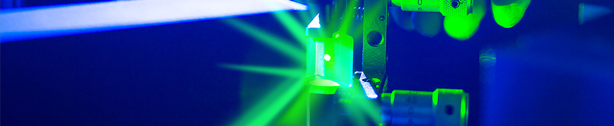
Feature: Surface Enhanced Raman Scattering in Biosensing & Material Analysis
Surface enhanced Raman scattering (SERS) is a technique which offers greatly improved Raman intensity for material analysis. Enhancement factors can be as high as 1012-13, which are sufficient to allow even single molecule detection using Raman. SERS is of interest for trace material analysis, flow cytometry and other applications where the current sensitivity/speed of a Raman measurement is insufficient.
The enhancement takes place at a metal surface which has nanoscale roughness, and it is molecules adsorbed onto that surface which can undergo enhancement. Typical metals used are gold or silver - preparation of the surface can be through electrochemical roughening, metallic coating of a nano-structured substrate, or deposition of metallic nanoparticles (often in a colloidal form). The SERS field has dramatically progressed from the originally observed enhancement on roughened silver electrodes to the current fields of sensing and imaging applications, single molecule detection, and extensions to ultrahigh vacuum and ultrafast science.
Performing a SERS experiment requires careful consideration of sample and optical setup to ensure maximum signal generation and enhancement. The first parameter is the enhancing substrate. Substrates range in structure from nano-rods to three-dimensional colloidal solutions with tunable plasmon resonances. Many researchers create their own SERS substrates, but commercially available kits offer a more routine approach. Next to consider is an appropriate excitation source, which should enable efficient excitation of the plasmon resonance. Typically it is necessary to use a laser wavelength which is compatible with the chosen SERS metal. Experimentally, it has been found that the maximum SERS signal is observed when the plasmon frequency is tuned to be slightly red-shifted from the laser wavelength.
Following excitation of the plasmon resonance and generation of the SERS signal, the detection process is identical to normal Raman experiments. A notch or long-pass filter is used to absorb or reflect any Rayleigh scattering while allowing for transmission of the Raman signal, and a spectrograph and detector are used to image Raman spectra across a wide spectral region.
Practically, the advantages of SERS can be explored on any Raman system, including commercial hand-hand Raman detectors. The power of SERS lies in its ability to identify chemical species and obtain structural information in a wide variety of fields including polymer and materials science, biochemistry and biosensing, catalysis, and electrochemistry. SERS biosensors are used in detection of various biological samples and diseases, including various cancers, Alzheimer's disease and Parkinson's disease, and recently also for glucose detection in Diabetes research.
Other applications of SERS method include toxic chemical detection, spectro-electrochemistry - the study of molecules in various oxidation states, art preservation, detection of counterfeit goods and currency, among others. Future directions for SERS holds promise in the fields of UV-SERS, Ultrafast and Stimulated SERS and Tip-Enhanced Raman Spectroscopy (TERS), which we will cover in a future Wavefront editon. Laserglow Technologies offers many laser wavelengths which are well suited for exploring standard and enhanced SERS experiments. We welcome the opportunity to collaborate with researchers in this field.
Sources:
1. "SERS: Materials, applications and the Future", Van Duyne et al. 2012, Department of Chemistry, North Western University Published Paper
2. SERS application notes- Horiba Scientific: Application Notes
3. "Principles of Surface-Enhanced Raman Spectroscopy" Eric C. Le Ru and Pablo G. Etchegoin ISBN: 978-0-444-52779-0
Laserglow in Research: Spectroscopy Method Explored for Water Safety
Laserglow's LRD-635 series 100 mW diode laser was recently used by researchers at San Diego State University in a study that aims to detect the presence of malachite green and crystal violet in a method based on nonlinear wave mixing and capillary electrophoresis. Although both of these compounds have antibacterial properties, they are reported to have toxic effects with time. This has lead the Food and Drug Administration (FDA) to label them as unsafe for medical use.
Due to health risks and the potential for illegal use in trace amounts, a very sensitive analytical detection method is needed to identify trace quantities in an aquaculture setting. Both malachite green and crystal violet are readily absorbed into fish tissues, which may then be ingested by consumers. Current detection methods often involve expensive, complicated techniques such as mass spectrometry to obtain low detection limits. What's required is a simple yet sensitive detection method that can be field deployed to confirm traces of these compounds in water supplies and animal tissues.
The detection method suggested by the researchers utilizes absorptivity, molecular weight and charge to identify these drugs at sub-nanomolar concentrations by laser wave mixing coupled with capillary electrophoresis (CE). Wave-mixing spectroscopy provides a sensitive absorption-based detection method for trace analytes. This is accomplished by forming dynamic gratings within a sample cell, which diffracts light to create a coherent laser-like signal beam with high optical efficiency and high signal-to-noise ratio. A cubic dependence on laser power and square dependence on analyte concentration make wave mixing sensitive enough to detect molecules in their native form without the use of fluorescent labels for signal enhancement.
In this method, a 532 nm laser and a 635 nm laser were used for malachite green and crystal violet sample excitation. The use of two lasers of different wavelengths allows the method to simultaneously detect both analytes. Selectivity is obtained through the capillary zone electrophoresis separation, which results in characteristic migration times. The method does not require any derivatization or labeling of the target analytes, and it is capable of producing reproducible results in less than two minutes. It yields picomolar detection limits in a detection system that can be scaled down to field-deployable designs.
The researchers suggest that further development of this method may offer even greater sensitivity and reproducibility. For a complete discussion of the results, go to: PubMed Full Paper
News: Theory Challenging Einstein's Speed of Light Could Soon be Tested
Scientists behind a theory that the speed of light is variable - and not constant as Einstein suggested - have made a prediction that could be tested. The assumption that the speed of light is constant, and always has been, underpins many theories in physics, such as Einstein's theory of general relativity. In particular, it plays a role in models of what happened in the very early universe, seconds after the Big Bang. But some researchers have suggested that the speed of light could have been much higher in this early universe.
Now, one of this theory's originators, Professor Joao Magueijo from Imperial College London, working with Dr Niayesh Afshordi at the Perimeter Institute in Canada, has made a prediction that could be used to test the theory's validity. In a paper published last week, he and Afshordi describe for the first time how scientists can finally test the controversial idea.
Magueijo and Afshordi first came up with a theory to explain why the cosmos looks much the same over vast distances. To be so uniform, light rays must have reached every corner of the cosmos, otherwise some regions would be cooler and denser than others. But even moving at 1bn km/h, light was not travelling fast enough to spread so far and even out the universe's temperature differences. To explain this dilemma, cosmologists including Stephen Hawking, proposed the theory of Inflation, which theorizes that the temperate of the cosmos evened out before the big bang. Magueijo and Afshordi's theory does away with inflation and replaces it with a variable speed of light.
According to their calculations, the heat of universe in its first moments was so intense that light and other particles moved at infinite speed. Under these conditions, light reached the most distant pockets of the universe and made it look as uniform as we see it today. The theory predicts a clear pattern in the density variations of the early universe, a feature measured by what is called the "spectral index".
Writing in the journal Physical Review, the scientists predict a very precise spectral index of 0.96478. Science can never prove the theory right. But Afshordi said that if measurements over the next five years shifted the spectral index away from their prediction, it would rule out their own theory. Read more at: Phys.org full article