x
Subject
Market Segment
Full Name
Company/Institution
Email
Phone
How Can We Help?
   Can We Help?
x
  Request Callback
Scientific Wavefront News & Blog

Integrating Light-Sheet Imaging With Virtual Reality To Recapitulate Developmental Cardiac Mechanics

Published 2017-11-28


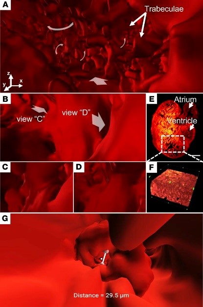
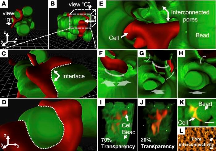
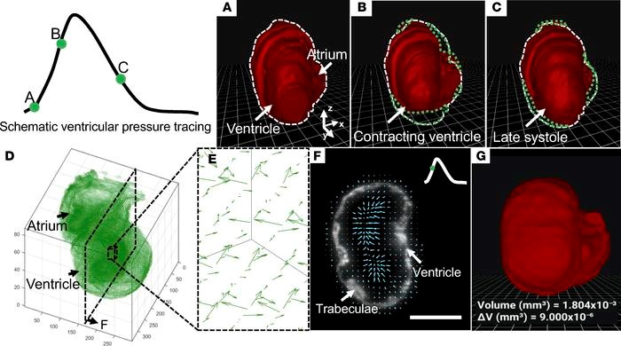
There is currently a limited ability to interactively study developmental cardiac mechanics and physiology. Researchers at UCLA combined light-sheet fluorescence microscopy (LSFM), constructed using continuous wave lasers from Laserglow, with virtual reality (VR) to provide a hybrid platform for 3D architecture and time-dependent cardiac contractile function characterization. By taking advantage of the rapid acquisition, high axial resolution, low phototoxicity, and high fidelity in 3D and 4D (3D spatial + 1D time or spectra), this VR-LSFM hybrid methodology enables interactive visualization and quantification otherwise not available by conventional methods, such as routine optical microscopes. They hereby demonstrate multiscale applicability of VR-LSFM to interrogate skin fibroblasts interacting with a hyaluronic acid-based hydrogel, navigate through the endocardial trabecular network during zebrafish development, and localize gene therapy-mediated potassium channel expression in adult murine hearts. They further combined their batch intensity normalized segmentation algorithm with deformable image registration to interface a VR environment with imaging computation for the analysis of cardiac contraction. Thus, the VR-LSFM hybrid platform demonstrates an efficient and robust framework for creating a user-directed microenvironment in which they uncovered developmental cardiac mechanics and physiology with high spatiotemporal resolution.


Design & Construction Of The Imaging System

The light-sheet imaging systems help to increase the working distance with sufficient spatial resolution needed to track organ development. The light-sheet microscope was built using continuous-wave laser as the illumination source. The detection module was installed perpendicular to the illumination plane, and it was composed of the scientific CMOS (ORCA-Flash4.0) and a set of filters (Semrock). During scanning, the sample holder was oriented by a 5-axis mounting stage. Both illumination and detection modules were controlled by a computer with dedicated solid-state drive redundant array of independent disks level 0 storage for fast data streaming.


Studies of neural pathways that contribute to loss and recovery of function following paralyzing spinal cord injury require devices for modulating and recording electrophysiological activity in specific neurons. These devices must be sufficiently flexible to match the low elastic modulus of neural tissue and to withstand repeated strains experienced by the spinal cord during normal movement. Inspired by preclinical and early clinical studies indicating the promise of spinal stimulation to facilitate rehabilitation following paralyzing injury, recent work has focused on flexible and stretchable probes for optical and electrical stimulation on the surface of rodent spinal cords.


Results

The VR-LSFM hybrid method establishes an efficient and robust platform to recapitulate cardiac mechanics and physiology. This framework builds on the high spatiotemporal resolution provided by LSFM to offer user-directed acquisition and visualization of life science phenomena, thereby facilitating biomedical research and learning via readily available smartphones and VR headsets. The application of BINS and DIR on LSFM-acquired data generates the input for a VR system, allowing for multiscale interactive use. The terminal platform composed of a smartphone and the Google Cardboard or Daydream viewer effectively reduces barriers to interpret the optical data, thereby paving the way for a virtual laboratory for discovering, learning, and teaching at single-cell resolution.


The VR-LSFM hybrid platform holds promises for the next generation of microscale interactions with live models at the cellular and material interface. Elucidating the dynamics of cardiac development and repair would accelerate the fields of developmental biology. The VR-LSFM platform demonstrates the capacity for improving the understanding of developmental cardiac mechanics and physiology with an interactive and immersive method.

To read more about this exciting research, please Click Here

MORE BLOG POSTS