
Can We Help You?
x
In this issue:
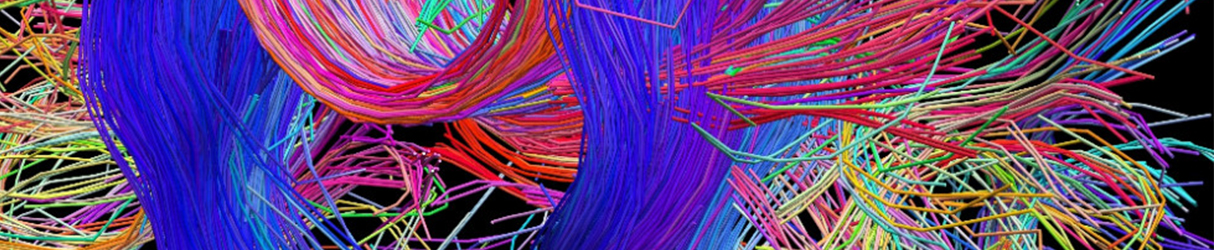
Clarifying the Human Connectome
In July of 2009, the National Institutes of Health (NIH), USA launched the Human Connectome Project aimed at building a map of the human brain, thereby shedding light on functional and anatomical connectivity of neurons, as well as to produce data that will facilitate research into brain disorders. Four years later in April 2013, the White House launched the BRAIN initiative (Brain Research through Advancing Innovative Neuro-technologies) as part of the Obama administration's broader Neuroscience Initiative. Both of these projects are part of the three 'Grand Challenges' of NIH's blueprint for Neuroscience research. As the International Year of Light draws to an end, we take a look at one of the methods that overlaps both the Connectome Project and BRAIN, and has shown promising results in understanding the science of memory, sleep and cognition.
The biggest challenge in understanding brain function is the complexity of the neural network. The human brain consists of around 86 billion neurons that interact with each other in complex circuits. Furthermore, the brain has a high lipid content, which makes it opaque and limits the depth of light penetration. Experiments that involve slicing the brain to understand its circuitry have their own limitations. The sectioned brain tissue may not capture the full extent of neuronal projections (which may extend hundreds of micrometers), thus providing an incomplete view of neuronal branching and regional variations in spine density. Thus, an ideal method to interrogate tissue is to microscopically examine it intact, rather than sectioned.
CLARITY is a technique that produces structurally intact, yet optically transparent brain tissue, which may be labeled and imaged without sectioning. The method was introduced in 2013 by Karl Deisseroth and his colleagues at Stanford University School of Medicine, and has since been implemented across many labs throughout the world. It involves applying a series of chemical treatments to a tissue sample in which the lipid content is removed from the cellular structure while almost all of the original proteins and nucleic acids are left in place, to achieve transparency. This enables microscopic study of the tissue's functional parts (predominantly proteins and nucleic acids).
CLARITY uses a strong detergent, SDS, to remove lipids from the tissue. To preserve protein integrity and the 3D structure of the tissue, it is first perfused with a formaldehyde and acrylamide solution that forms structural hydrogel scaffolding. With the aid of an electrophoretic current SDS then removes the lipids that are not linked to the scaffold. Cleared tissue may then be labeled and/or imaged with substantially increased light penetration and high-resolution images may be collected several millimeters or more below the tissue surface.
Subsequent studies using the CLARITY method have included studies on Alzheimer's diseased human brains, mouse spinal cords, multiple sclerosis animal models, and microscopy methods including tissue expansion or swelling for use in confocal microscopy (F. Chen et al., Science, Jan. 2015) and CLARITY-enhanced light sheet microscopy or COLM. Optimization of CLARITY technique involves varying a number of factors - like temperature, hydrogel composition, polymerization conditions and lipid-clearing methods to provide a stable and replicable procedure for rendering tissues transparent.
Sources:
1. 'Optimization of CLARITY for Clearing Whole-Brain and Other Intact Organs' - K. Deisseroth et al., ENEURO: Online Link-ENEURO
2. 'An Illuminated State of Mind'- Marie Freebody: Online Link-Photonics.com
3. 'See-through brains clarify connections'- Helen Shen: Online Link-Nature News
4. 'Brain Research through Advancing Innovative Neurotechnologies' - The White House BRAIN Initiative: Online Link-BRAIN
Tougher Collagen Hydrogels Enable Safe Delivery of Healthy Cells into Joints
A team of researchers at Harvard Medical School in collaboration with University Medical Center, Utrecht have proposed a method that enhances the stiffness of collagen gel- primarily used as a scaffolding in cartilage tissue engineering, to safely deliver healthy chondrocyte cells into joints during surgery. Laserglow's LRS-0532 LabSpec laser was used as a light source.
Cartilage doesn't naturally heal on its own. It is populated exclusively by chondrocytes (the only cells found in a healthy cartilage), which produce and maintain the cartilaginous matrix that consists of collagen and proteins. Any procedure aimed at cartilage regeneration aims at creating a natural hyaline cartilage to fill the defect, and allowing integration of the neo-cartilage with existing healthy cartilage. Collagen naturally occurs in the human body so it's a good choice for a cartilage scaffolding. However, in its natural state it lacks mechanical integrity and is easily digested by enzymes.
Exposing collagen gels containing photo reactive dyes to green laser light induces cross-links to adjacent gel molecules. The chondrocytes encapsulated in photo-crosslinked collagen hydrogels formed hyaline-like cartilage. To strengthen the resulting hydrogels, the researchers then treated them with a 1 hour exposure to EDC and NHS- both common chemical activating agents. The researchers found that this treatment more than doubled the collagen's resistance to enzyme attack, and increased its storage modulus five-fold. The researchers are confident that this procedure will greatly increase the effectiveness of in-vivo cartilage regenerative efforts.
The research was first published online on 22 July 2014 in Wiley Online Library, DOI: 10.1002/jbm.a.35266. To learn more about the experimental setup and materials used, read the full paper at: Wiley Online Library Full Paper
Canadian Laser System Begins Voyage to Explore Asteroid
A laser system developed by researchers at York University and built in collaboration with several Canadian companies is set to explore a near-earth asteroid named Bennu. The system is Canada's contribution to a NASA-led mission called OSIRIS-REx that will try to collect samples from Bennu at its closest point to earth. Scientists hope the mission will reveal clues to the early history of the solar system, as well as provide groundwork for strategies on how to divert objects that end up on a collision course with our planet. Bennu currently has a 1 in 2700 chance of colliding with earth in the next century.
The Canadian component of the mission is known as OLA (short for OSIRIS-REx Laser Altimeter). Once deployed, it will be essential for mapping the asteroid's irregular surface and helping mission scientists select locations where the spacecraft can collect a sample for analysis. OLA is currently at Lockheed Martin's Denver facility, where the main OSIRIS-REx spacecraft is in the final stages of assembly and testing. The spacecraft is set to launch in September 2016, and is scheduled to rendezvous with Bennu in August 2018.
Normally, a laser altimeter is a stationary device that uses laser light to measure the distance from an orbiting spacecraft to the surface below. After many orbits, this data can be assembled into a topographical map. What makes OLA different, and more ambitious, is the addition of a scanning mirror that allows the laser beam - pulsing at 10,000 times per second (10 kHz) - to create an accurate three-dimensional representation of an asteroid's surface with near-photographic speed. This will provide valuable clues on Bennu's composition and history to scientists, and will also help guide the spacecraft when looking for a suitable location for sample collection. Read more at: Globe and Mail full article