
Can We Help You?
x

In this issue:
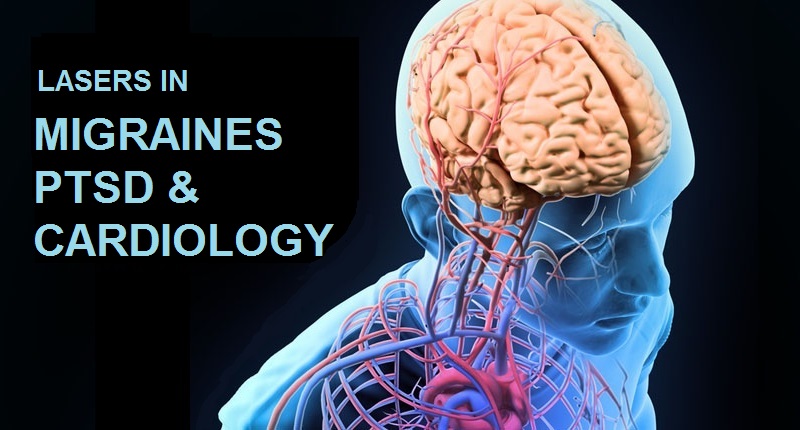
Latest in Optogenetics: Migraines, PTSD and Cardiology. Plus, Cyanea in the lab!
The World Economic Forum recently named Optogenetics as one of the top 10 emerging technologies of 2016. Several industry reports have suggested that the growth of this technique in biotech investigations, especially in relation to neuroscience will increase fivefold in the next decade. In this feature, we take a look at some of the latest published research that has employed Optogenetics as the primary investigative technique.
As with any neuroscience related investigation technique, one fundamental challenge facing optogeneticists is being able stimulate a small selection of neurons, without causing any peripheral damage to neighboring neurons. Now, ultrathin, flexible microchips, each one hardly bigger than a neuron, are being tested as injectable devices to put selected nerves under appropriately small wireless control. These microchips can be inserted deep into the brain with minimal damage to overlying tissue. Researchers at the Washington University School of Medicine, St. Louis, and University of Illinois, Urbana-Champaign, created a remote controlled tissue implant that allows neuroscientists to inject drugs and shine light on neurons deep inside the brain of the subject. With a thickness of 80 micrometers and a width of 500 micrometers, the opto-fluidic implant is thinner than the metal tubes, or cannulas, that scientists typically use to inject drugs. The scientists tested the device's drug delivery potential by surgically placing it into the brains of mice. In some experiments, they showed that they could precisely map circuits by using the implant to inject viruses which label cells with genetic dyes. In other experiments, they made mice walk in circles by injecting a drug that mimics morphine into the ventral tegmental area (VTA), a region that controls motivation (and addiction).
In another study, researchers at the Icahn School of Medicine at Mount Sinai, New York, NY, used a combination approach that included optogenetics to discover that the process responsible for persistent reactions to fear-associated cues is influenced by a sparse but potent population of inhibitory cells called parvalbumin-interneurons (PV-INs) in the amygdala. They found that when a specimen acquires a fear memory, the suppressive influence of PV-INs is relieved, allowing the fear system to respond more vigorously when the auditory stimulus is re-encountered to trigger a 'fight-or-flight' response. Roger Clem, PhD., the principal investigator of this study believes that further research into PV-INs could lead to clues on how to structure therapy and treatments for PTSD (Post Traumatic Stress Disorder).
In an article recently published in Current Neuropharmacology, Feng Tao, MD, PhD. associate professor in the Department of Biomedical Sciences at the Texas A & M University College of Dentistry discusses how optogenetic manipulation can also be used for the study and treatment of pain, including migraine-related pain. Targeting neural networks involved in migraine pathophysiology, this technology shows great promise to reduce migraine-related headaches. "This technology won't just be used for biomedical research," Tao said in a statement. "We hope we will be able to move it into clinical trials to treat intractable pain."
Lastly, an optogenetic technique has been used to make cardiac cells beat and optically measure their response, enabling an automated drug-testing process called OptoDyCE. The fully automated system for all-optical cardiac electrophysiology can rule out potentially dangerous drugs by testing 30,000 light-responsive cells in less than 10 minutes, potentially replacing a process that currently takes hours or even years. The study was conducted by Professor Emilia Entcheva's lab at George Washington University, and first published in Nature Communications.
As many of you may know 470 nm is the peak response wavelength for the broadest use of optogenetics, that of Channelrhodopsin (and associated) opsins. We conclude this feature with a promising update on the Cyanea Series of 470 nm blue diode laser which Laserglow launched last month. A comprehensive series of benchtop tests of the laser unit were conducted at Concordia University, Montreal, QC, Canada and this objective set of data confirms the greatly improved performance of this new system. From near cold starts up to operating temperature, and with modest to very low duty cycles, as well as short pulse trains to elongated events, the Cyanea system outperformed the 473 nm DPSS system with incredible ease. The initial reports of these performance benefits are very favorable and confirm the superiority of this collimated laser diode system over the DPSS units that have been used extensively around the globe for many years. We invite the academic community to contact us at [email protected] to view the detailed performance report. Laserglow is very grateful to the Concordia team for sharing their results.
Research Update: MRI-Guided Focal Laser Therapy for Prostate Cancer
A Laserglow 671 nm LabSpec™ laser was recently used as a light source in a study using MRI-Guided Focal Photothermal Therapy (PTT) to destroy tumorous prostate cancer cells. The research was conducted at the Princess Margaret Cancer Center in Toronto, Canada in collaboration with University of Toronto and Peking University, China.
In the past, MRI-Guided Focal Photothermal laser therapy has been investigated during phase 1 trials of treating intermediate risk localized prostate cancer cells. The progress however, has been limited due to lack of tumor selectivity and low efficiency of heat generation (the thermal aspect of using the laser). In this study, intrinsic multi-functional porphyrin nanoparticles (porphysomes) have been exploited to treat localized prostate cancer by MRI-Guided Focal Photothermal Therapy with significantly improved efficiency. The PTT was conducted with MRI-guided light delivery and monitored by MR thermometry, mimicking the clinical protocol. The efficacy of treatment and adverse effects to surrounding tissues were evaluated by histology analysis and tumor growth in survival study (via collected MRI data).
The researchers found that Porphysomes showed superior tumor-to-prostate selectivity over current clinical standards. MR thermometry detected tumor temperature increasing to ≥55° °C within 2 minutes when irradiated by the 671 nm wavelength laser at 500 mW, yet showed minimal thermal increases in surrounding tissues. Porphysome enabled effective PTT eradication of the tumor without damaging adjacent organs in the orthotropic prostate cancer specimen.
The researchers believe that MRI-guided focal PTT could be an effective and safe approach to treat prostate cancer with low risk of progression, thus addressing the significant unmet clinical needs while benefiting an ever-growing number of patients who may be over-treated and endure risk of unnecessary side effects from more radical therapies. For the complete paper and detailed discussion of experimental setup go to: PubMed Full Paper
Updated Map of the Human Brain Shows 180 Different Regions
A group of researchers at Washington University have updated the 100-year-old map of the human brain (originally made by German neurologist Korbinian Brodmann) in a scientific feat which revealed that the human brain has at least 180 different regions individually and cumulatively important for language, perception, consciousness, thought, attention and sensation. This discovery confirms the existence of the previously known 83 regions, while adding 97 new ones.
The landmark achievement hands neuroscientists their most comprehensive map of the cortex so far, one that is expected to supersede Brodmann's as the research standard to be used when discussing the various areas of study or examination of the brain. The researchers created the map by combining highly-detailed MRI scans from 210 healthy young adults who had agreed to take part in the Human Connectome Project, which is a globally engaged, massive multi-discipline and multi-institute effort that aims to understand how neurons in the brain are connected.
Writing in the journal Nature, Matthew Glasser and others describe how they combined scans of brain structure, function and connectivity to produce the new brain map. Some scans were taken while patients simply rested in the test equipment, while others were recorded as they performed math tasks, listened to stories, or categorized objects, (for example by stating whether an image was of a tool or an animal).
The detailed brain map, which will be made freely available for all, should help scientists working in the field of neuroimaging be more certain about the areas of the brain in which they see activity or notice problems within, when scanning patients and volunteers. The map will have an immediate impact on fundamental brain research, but will also quickly be taken up by neurosurgeons who can use the scientists'computer algorithm to identify all of the different brain regions in patients they are about to operate upon. Read more at: The Guardian full article