In this issue:

Advancements in Biomedicine through Laser Ablation Techniques
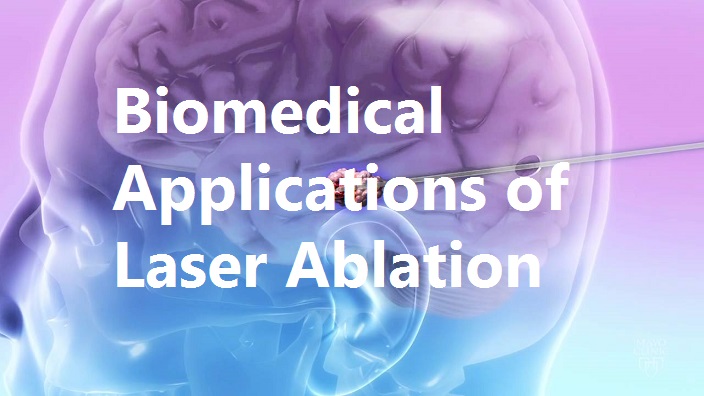
Laser Ablation is the process of removing material from a surface by irradiating it with a laser beam. Laser-based ablation is greatly affected by the nature of the material and its ability to absorb energy, therefore the wavelength of the ablation laser should have a minimum absorption depth. While mostly this term references pulsed lasers, it's also possible to ablate a surface using a Continuous Wave laser beam if the intensity and duration of use is optimized. Within biological sciences, laser ablation has been used to study cell function, cell structure, tissue interaction, gene structuring, and neuron interaction and is now increasingly used within medicinal fields such ophthalmology, dermatology, oncology and neurosurgery.
The selective laser cutting of cellular structures or the removal of whole cells by ablation allow the study of cell and developmental biology. This technique provides new insights into the process of chromosome segregation as well as cell locomotion. Furthermore, it gives new information about origin, fate, or function of individual cells in the developing organism and can also be used for gene activation. One primary advantage of laser ablation in cell biology is the flexibility of the method, which can be performed at any cell site, and at any time in the development within living tissue and cells. When pulsed lasers are coupled to a laser-scanning microscope for selective photo-modulation, it becomes possible to target specific cell organelles. Efficient ablation requires pulsed laser excitation to reach high peak intensities with moderate average power. In this way, adjacent cell damage is minimized in untreated regions. High energy, localized pulses allow control over the heat generation in the tissue which is significantly reduced due to the short pulse duration.
Recently, laser ablation inductively coupled plasma mass spectrometry (LA-ICP-MS) has widely been used for the analysis of biological samples. The technique allows the determination of elements and isotopes in biological tissues and related materials with a spatial resolution typically ranging from 10 to 100 microns. This eases the detection of metals and metalloids in tissues through bio-imaging. Laser enabled thermal ablation is also used for removal and reduction of tumors, specifically solid neoplasms. Many experimental studies have shown progress of cell-tumor death following ablation, and this method is now combined with immunotherapy, conventional chemotherapy and radiation to treat tumors.
Within medicine, the last 2 years have seen a surge in the direct application of laser ablation in treatment of various diseases. In June 2015, data from the Sperling Prostate Center and the NYU School of Medicine (New York, NY) indicated that MRI-guided targeted laser ablation was 96% effective in destroying cancerous cells with almost no side-effects on neighboring organs. Laser ablation is also now being used in minimally invasive neurosurgery for patients with epilepsy involving highly localized seizures occurring in the hippocampus. In March 2016, the FDA approved a study to evaluate this approach in treating medically refractory epilepsy. And finally, in Feb 2016, researchers at Florida Atlantic University reported that a method involving Raman Spectroscopy following laser ablation that enabled them to differentiate skin cancer cells from regular skin cells.
While these are just a few of the biomedical applications that have benefited from development of laser ablation, there are several studies in place to explore this method for treatments that are currently painful, costly and / or have severe side effects. We can only expect to hear more about the benefits of laser ablation in biomedicine in the years to come.
Optogenetics Investigated for Treatment of Chronic Pain
Researchers at McGill University in Montreal, Canada have demonstrated the potential role of optogenetics in pain management through a study in which light-sensitive neurons were inhibited in the presence of laser light. A Laserglow 473 nm DPSS laser was the laser light source for fibre delivered optical stimulation of neurons.
The study is based on the premise that by making the cells responsible for pain transmission sensitive to light, it will be possible to target, desensitize and reduce bioelectric activity in these cells. The peripheral neurons that are known to transmit pain are known as nociceptors. The nociceptors of the subject in this experimental study expressed proteins called opsins that react to light. When the nociceptors were exposed to yellow light, the opsins moved ions across the membrane, reducing the level of bioelectric activity of the cells. The yellow light stimulation reliably blocked electrically induced action potentials in dorsal root ganglion (DRG) neurons in the subject, effectively shutting off the nociceptors and decreasing sensitivity to touch and heat.
The precision of this technique underlines potential advantages for use of a future version of this technique in humans. The researchers believe that light therapy based on optogenetics would have the advantage of providing on-demand analgesia to patients who could control their pain by shining light on the sensitive part of the body. To learn more about how blue and yellow laser lights were used together in this study, and for detailed discussion of results, read the full paper at eNeuro Full Paper
Brightest X-ray Laser Vaporizes Water - Video Released
Scientists have recorded the first ever microscopic movies of water being vaporized by the world's brightest X-ray laser. The data gathered at the Department of Energy's SLAC National Accelerator Laboratory, in Menlo Park, California, could shed new light on X-ray lasers, and how these extremely bright, fast flashes of light take atomic-level snapshots of some of nature's speediest processes.
"Understanding the dynamics of these explosions will allow us to avoid their unwanted effects on samples," says Claudiu Stan of Stanford PULSE Institute, a joint institute of Stanford University and SLAC. "It could also help us find new ways of using explosions caused by X-rays to trigger changes in samples and study matter under extreme conditions. These studies could help us better understand a wide range of phenomena in X-ray science and other applications."
The team injected water into the path of the laser as a series of individual drops, as well as a continuous jet. As each individual X-ray pulse hit the water, a single image was recorded, timed from five billionths of a second to one ten-thousandth of a second after the pulse. These images were then strung together to create the movies.
Liquids are commonly used to put scientific samples into the path of an X-ray beam for analysis. The experiments show in detail how the explosive interaction unfolds and provides clues as to how it could affect X-ray laser experiments. The study was published this week in the journal Nature Physics. Read more and watch the video at Phys.org full article