In this issue:

Traumatic injuries to the spinal cord are frequently associated with loss of organ function or loss of voluntary limb control. Our understanding of and ability to treat these symptoms is currently limited by the tools available for monitoring and manipulating neural dynamics within the spinal cord.
But now, researchers focusing in the field of
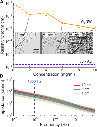
Optogenetics at MIT, have been able to confirm the ability of endogenous electrophysiological activity in the spinal cord of transgenic mice. Because of the relative ease of genetic manipulation, rodent models have become indispensable discovery tools for basic neuroscience. They report using flexible, stretchable probes consisting of thermally drawn polymer fibers coated with micrometer-thick conductive meshes of silver nanowires. Simultaneous stimulation and recording was demonstrated in transgenic mice expressing channelrhodopsin 2 (ChR2), where optical excitation, using Laserglow's
473nm Blue DPSS Laser, evoked electromyographic activity and hindlimb movement correlated to local field potentials measured in the spinal cord.
Studies of neural pathways that contribute to loss and recovery of function following paralyzing spinal cord injury require devices for modulating and recording electrophysiological activity in specific neurons. These devices must be sufficiently flexible to match the low elastic modulus of neural tissue and to withstand repeated strains experienced by the spinal cord during normal movement. Inspired by preclinical and early clinical studies indicating the promise of spinal stimulation to facilitate rehabilitation following paralyzing injury, recent work has focused on flexible and stretchable probes for optical and electrical stimulation on the surface of rodent spinal cords.
Electrophysiological recording of spontaneous, sensory-evoked, and optically evoked neural activity in the spinal cords of mice expressing channelrhodopsin 2 (ChR2) further illustrates the promise of these hybrid fiber probes for studies of spinal cord circuits. Recording of endogenous activity in freely moving mice chronically implanted with fiber probes in their spinal cords, combined with analysis of the surrounding tissue, suggests minimal disruption to local neural networks.
These findings suggest that the fiber platform may, in the future, permit monitoring and controlling of neural activity to promote recovery following spinal cord injury. Isolating single-neuron action potentials from the mouse spinal cord during free behavior remains a goal because such recordings are exceedingly difficult, having been rarely reported in rodents . Even in acute studies, the flexibility of the probes may provide a solution to the challenges associated with the recording of neural activity in the spinal cord posed by the respiration and heartbeat, which often necessitate a transection of nerves leading to the diaphragm.
To read more about this exciting research, please -
Click Here
To learn more about the laser used in the research, please visit -
Click Here

