
Can We Help You?
x
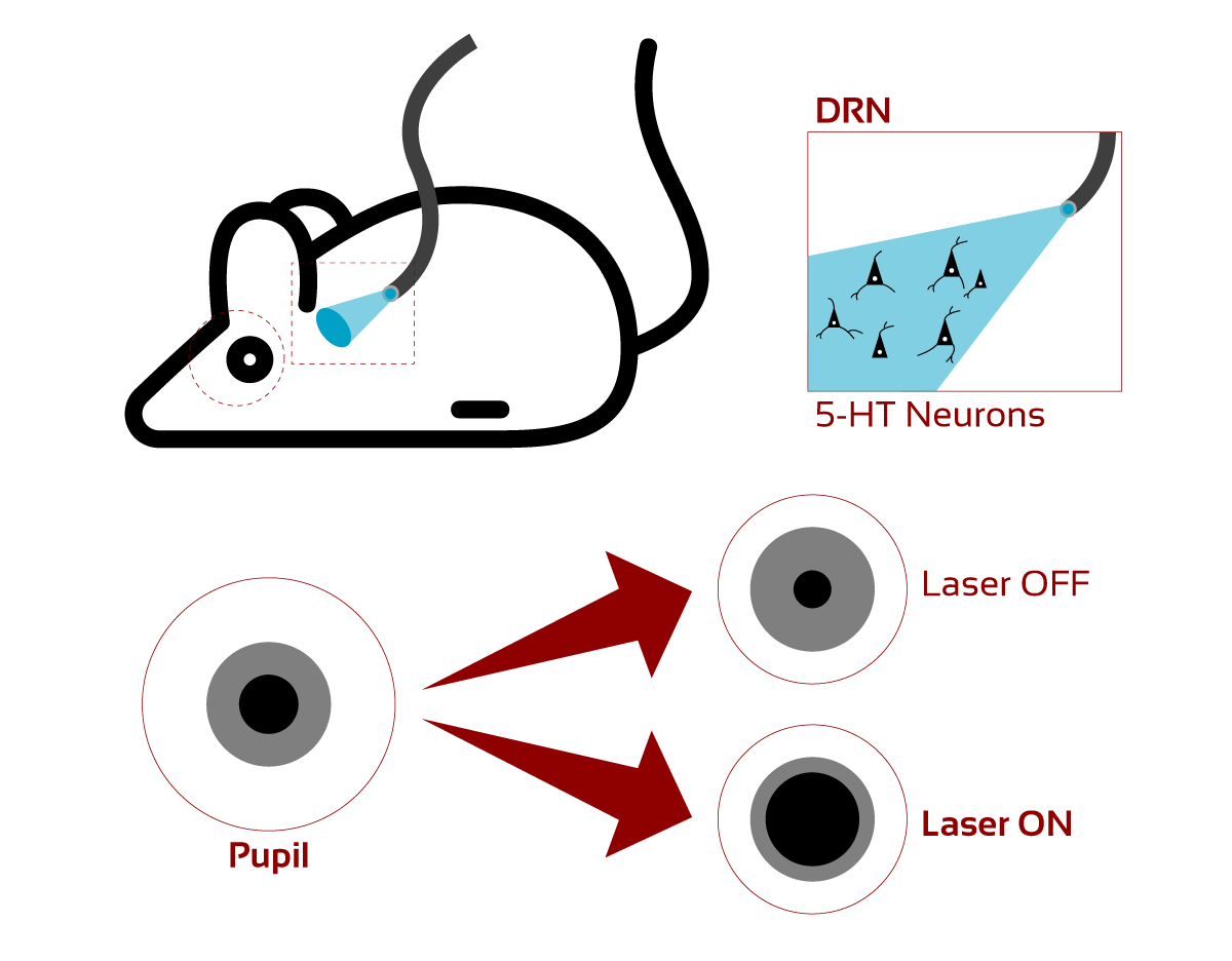
Transient variations in pupil size under constant luminance are coupled to rapid changes in arousal state, which have been interpreted as vigilance, salience, or a surprise signal. Neural control of such fluctuations presumably involves multiple brain regions and neuromodulatory systems, but it is often associated with phasic activity of the noradrenergic system. Serotonin, a neuromodulator also implicated in aspects of arousal such as sleep-wake transitions, motivational state regulation, and signaling of unexpected events, seems to affect pupil size, but these effects have not been investigated in detail. In this study, researchers from Champalimaud Centre for the Unknown (Lisbon, Portugal) show that phasic serotonin neuron stimulation causes transient pupil size changes. The researchers used optogenetic activation of serotonin neurons in the dorsal raphe nucleus of head-fixed mice performing a foraging task. Serotonin-driven modulations of pupil size were maintained throughout the photostimulation period and sustained for a few seconds after the end of stimulation. The researchers found no evidence that the increase in pupil size with activation of serotonin neurons resulted from interactions of photostimulation with behavioral variables, such as locomotion or licking. Furthermore, the researchers observed that the effect of serotonin on pupil size depended on the level of environmental uncertainty, consistent with the idea that serotonin could report a surprise signal. These results advance our understanding of the neuromodulatory control of pupil size, revealing a tight relationship between phasic activation of serotonin neurons and changes in pupil size.
Methods: Optogenetic Stimulation
To stimulate optically ChR2-expressing serotonin neurons, the researchers used a laser emitting blue light at 473 nm (Laserglow’s LRS-0473-PFF-00800-03). Light was emitted from the laser through a rotary joint via optical fiber patch-cords, to the chronically implanted optic fiber cannula. Optical stimulation was exclusively turned on during 30% of short sessions, only during their foraging task - after the first lick was detected, up to 5 s unless the animal moved interrupting the stimulation.
Previous studies recorded dorsal raphe nucleus electrophysiological responses in mice expressing ChR2 in the dorsal raphe nucleus while optogenetically stimulating the dorsal raphe nucleus neurons using parameters like this study. This demonstrates that Serotonin neurons reliably respond to the stimulation protocol throughout the duration of the pulse and did not detect evidence that the effects of stimulation fade away over time. Additional studies show that increasing the pulse frequencies of the photostimulation produces a monotonically increasing dose-dependent effect, so the researchers used the pulse frequency that yields the largest behavioral response These studies, along with others, provide strong evidence that the optogenetic approach yields selective and effective activation of Serotonin neurons.
Click here to access to the materials and methodology.
Click here to learn more about the 473 nm laser used in the research.