
Can We Help You?
x
What is Fiber Photometry?
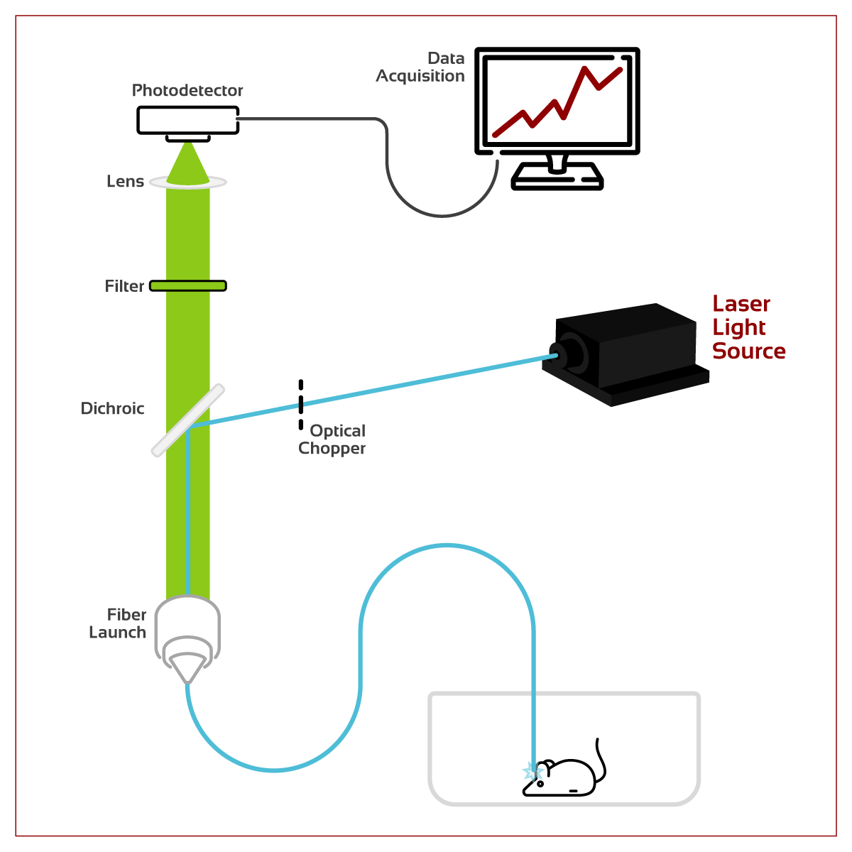
Fiber Photometry is a neuroscience research method that optically records in real-time natural, specific-connected neutral activity by projecting light down optical fiber into a live rodent via an implant. This light can be used for activating optogenetic actuators and for exciting injected or genetically encoded reporters, typically Calcium indicators. Light emitted from these reporters can be transmitted back along the same fiber and separated from the excitation light with dichroic mirrors before detection by a photodetector set up.
Advantages
Fiber photometry is an easy-to-use and economical system – the most basic setup allows the recording from a population of neurons without the additional complication of spatial information. Light sources and optical accessories are widely available and standardized, and compatible with most equipment commonly available in neuroscience laboratories, such as those with optogenetics setups.
A basic fiber photometry setup is simple and cost-effective, comprising of a laser light source, optical fiber, dichroic mirrors, bandpass filters, and a detector – making budget friendly star- up option or integration/modification to an existing optogenetics setup. During the recording interval, subjects are freely behaving, and multiple color channels can be simultaneously recorded using the same fiber. There are no electronics mounted to the head of the animal, therefore weight load is minimal and no heat is generated near the tissue.
Popular photodetectors are photodiodes and photomultiplier tubes (PMTs), both of which are sensitive to even single photon count levels, especially PMTs. Since these signals are picked up from a large volume, fiber photometry can detect even low activity levels. Advanced variations of more complex setups of fiber photometry also exist, such as providing increased channel counts, using cameras instead of a photodetectors, adding spectral unmixing, or customizing optical fibers to achieve optical and geometric qualities. With proper configuration, photodetectors are fast enough to detect action potentials, and take advantage in recent advancements in genetically encoded voltage indicators (GEVIs) to follow action potentials in fast-spiking neurons.
Disadvantages
Unfortunately, fiber photometry lacks spatial resolution because the recorded signal is an accumulation of fluorescence intensity from activity of the entire population. However, two-photon microscopy is an option that reduces the spatial resolution drawback of fiber-photometry and can record neuronal activity with sub-cellular resolution and great signal-to-noise ratio.
Wavelengths
Common wavelengths and their related applications in fiber photometry include:
Violet Light - 405 nm
Autofluorescence Detection: Autofluorescence (AF) is the emission of light from either brain tissue when excitation light is absorbed, or from the fiber optic components. While recording, AF increases with overall background noise resulting in a need for detecting and removing AF with appropriate optical fibers and techniques such as photo bleaching (permanently altering fluorophores using intense light exposure to make it unable to fluoresce).
Isosbestic Control: This is the control signal used for photobleaching and motion artifact. An isosbestic point is a specific wavelength at which total absorbance of a sample does not change during a chemical reaction or physical change. One can disambiguate signal from noise as using the isosbestic point of the commonly used GCaMP (a genetically encoded Ca2+ indicator). When GCaMP is excited at this point, its emission is unaffected by changes in internal calcium concentrations, and variance in its signal would indicate bending of optic fibres or movement of neural tissue.
Blue Light - 470/473 nm
GCaMP is a green fluorescent genetically encoded calcium indicator (GECI) commonly used in calcium imaging experiments. There are also recent generations optimizing for speed, signal-to-noise ratio, expression, and changes in fluorescence. Another example of a blue GECI would be dLight.
Yellow-Red Light - 561/589
Although blue GECIs are the most common, there are also yellow/orange/red options available, such as mCherry, tdTomatoc, RCaMP and XCaMP.
| Product | Wavelength | Description | Starting At | In Stock | |
|---|---|---|---|---|---|
 | 405 nm Best Seller! | 405 nm Collimated Diode Laser System 50 - 10000 mW Output Power | $991.00 | Specs |
|
 | 470 nm Best Seller! | 470 nm Collimated Diode Laser System 100 - 1500 mWOutput Power | $3,616.00 | Specs |
|
 | 473 nm Best Seller! | 473 nm DPSS Laser System 5 - 100 mW Output Power | $1,664.00 | Specs |
|
 | 561 nm | Fanless 561 nm DPSS Laser System 5 - 200 mW Output Power | $3,600.00 | Specs |
|
 | 589 nm Best Seller! | 589 nm DPSS Laser System 10 - 50 mW Output Power | $5,913.00 | Specs |